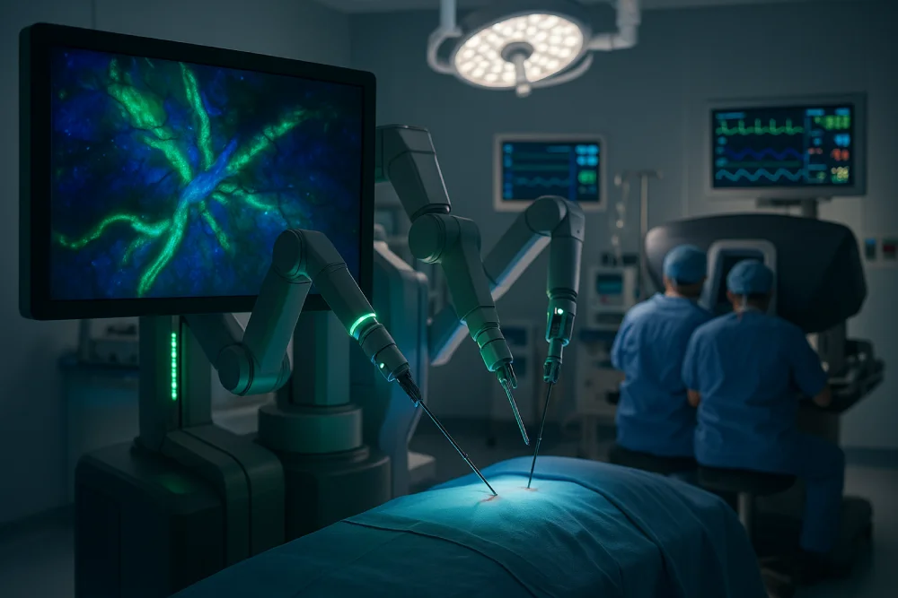
Firefly imaging technology maximizes robotic surgery precision by providing real-time near-infrared fluorescence visualization that reveals vascular perfusion and microvasculature invisible to standard cameras. This technology enables surgeons to differentiate between healthy and abnormal tissue, assess tumor margins accurately, and identify critical anatomical structures during complex procedures. Seamlessly integrated with Da Vinci robotic systems, Firefly enhances surgical outcomes across oncologic and reconstructive procedures while reducing complications and operative times. Advanced applications continue expanding across multiple surgical specialties.
How does firefly imaging alter the surgeon’s ability to visualize key structures during robotic procedures? Near-infrared fluorescence technology fundamentally transforms surgical visualization by revealing vascular perfusion and microvasculature invisible to standard cameras. When indocyanine green dye is injected, it fluoresces bright green under NIR light, enabling real-time identification of blood supply patterns and essential anatomical structures like ureters and bile ducts. This enhanced visualization provides clear differentiation between healthy and abnormal tissue, particularly important during tumor resection where margins become distinctly visible. However, dosage optimization remains pivotal for effective imaging, as improper amounts reduce accuracy. Dye limitations include variable performance in obese patients and post-radiotherapy cases due to altered distribution patterns, requiring surgeon experience for optimal interpretation. The technology’s magnified 3D visualization capabilities allow surgeons to examine anatomical structures with unprecedented detail and depth perception during complex reconstructive procedures.
Firefly imaging technology has demonstrated significant clinical utility across multiple surgical specialties, with particularly notable applications in oncologic and reconstructive procedures. In oncologic surgery, the technology enables precise differentiation between cancerous and normal tissue while facilitating accurate tumor margin assessment and lymphatic mapping across urologic, gynecologic, and colorectal specialties. Reconstructive procedures benefit from real-time perfusion assessment and enhanced anatomical visualization, allowing surgeons to evaluate tissue viability and optimize surgical outcomes in complex cases. The system’s organ transillumination capabilities provide enhanced visualization of anatomical structures during robotic gynecologic procedures, contributing to improved surgical precision and patient outcomes.
Precision in cancer surgery has reached new heights with the integration of near-infrared fluorescence imaging technology into robotic surgical platforms. Firefly imaging revolutionizes oncologic procedures by enhancing tumor margin detection, enabling surgeons to distinguish malignant tissue from healthy structures with unparalleled clarity. The technology employs fluorescent tracer research developments that illuminate cancerous cells in real-time, significantly improving surgical outcomes.
Sentinel lymph node identification becomes markedly more accurate, allowing precise assessment of cancer spread patterns. Vascular visualization capabilities help preserve critical blood vessels during complex tumor resections. The system provides immediate perfusion feedback, ensuring tissue viability throughout procedures. The da Vinci Xi system introduced enhanced illumination capabilities that further improved fluorescence visualization quality in oncologic applications. This minimally invasive approach reduces patient recovery time while maximizing surgical precision, fundamentally altering how oncologic surgeries are performed across multiple cancer types.
While oncologic applications have demonstrated significant advantages, reconstructive surgery represents another frontier where near-infrared fluorescence imaging alters surgical outcomes across multiple specialties. Firefly technology provides real-time, magnified 3D visualization of microperfusion and vascular supply during complex reconstructions. Surgeons can toggle between near-infrared and standard white light views to delineate healthy from diseased tissue, particularly meaningful in scarred surgical fields. Enhanced surgical ergonomics result from improved visualization without additional instrument manipulation. In ureteral reconstruction, Firefly enables precise tracking through disrupted tissue planes, minimizing inadvertent injury. The bright green cut ends indicate healthy tissue boundaries, allowing surgeons to identify sections requiring removal while minimizing future scar tissue formation. Perioperative workflow optimization occurs through reduced operative complications and shorter recovery times, with clinical series demonstrating average 5.9-day hospital stays and minimal postoperative morbidity across urology, gynecology, and general surgery applications.
Firefly imaging technology in robotic surgery directly translates to measurable improvements in patient outcomes through accelerated recovery timelines and enhanced procedural safety. The minimally invasive nature of robot-assisted procedures combined with precise fluorescence-guided visualization reduces tissue trauma, leading to shorter hospital stays and faster return to normal activities. This enhanced surgical precision significantly decreases both intraoperative and postoperative complications, resulting in improved overall patient safety profiles across multiple surgical specialties.
Among the most significant advantages of robotic surgery with Firefly imaging is the substantial reduction in patient recovery time compared to traditional surgical approaches. The minimally invasive nature of robotic procedures creates smaller incisions and causes less tissue trauma, resulting in reduced anesthesia requirements and shorter hospital stays averaging 5.9 days. Expedited mobilization becomes possible as patients can begin gentle movement shortly after surgery, reducing complications like profound vein thrombosis while accelerating overall healing. Enhanced precision through real-time visualization minimizes damage to healthy tissues and preserves critical anatomical structures. This careful tissue preservation correlates with decreased inflammatory response and reduced post-operative pain, allowing patients to resume basic daily activities within 3-7 days and rely on simple over-the-counter pain medications for comfort management.
Precision emerges as the cornerstone of reduced surgical complications when Firefly imaging technology integrates with robotic surgical platforms. The enhanced visualization capabilities enable surgeons to identify critical anatomical structures with unparalleled clarity, significantly reducing inadvertent injury to blood vessels and organs during complex procedures.
| Complication Reduction | Traditional Surgery | Firefly-Enhanced |
| Intraoperative Injuries | Higher risk | Minimized risk |
| Conversion to Open | Variable rates | Zero conversions* |
| Grade 2+ Complications | Standard rates | 12% (3/25 patients)* |
Real-time fluorescence imaging provides objective guidance that minimizes operator-dependent variability, leading to more consistent outcomes across surgical teams. This standardization particularly benefits centers with developing skill, where improved patient safety becomes achievable through enhanced visualization protocols that reduce reliance on subjective surgical judgment.
*Based on 25-patient series
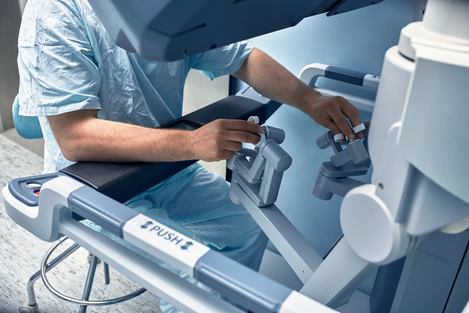
The seamless integration of fluorescence imaging technology into the da Vinci Surgical System represents a significant advancement in robotic surgical capabilities. Firefly’s modular design features enable direct embedding into da Vinci Xi and X systems without requiring additional hardware components or surgical instruments. The near-infrared fluorescence camera integrates with standard 3D endoscopic optics, allowing surgeons to toggle between visible light and fluorescence modes through console interface controls. This simplified workflow integration maintains the robotic surgical process while providing real-time fluorescence visualization. Specialized optical sensors and filtered endoscopes detect indocyanine green fluorescence, co-registering white light and fluorescence images on a single console screen. The system operates without extra ports or staff adjustments, preserving the streamlined nature of robotic surgery while enhancing intraoperative decision-making capabilities.
Building upon the sturdy hardware foundation of da Vinci integration, Firefly imaging alters surgical visualization through sophisticated vascular assessment and tissue differentiation capabilities. Near-infrared fluorescence technology employs indocyanine green dye to provide real-time magnified 3D visualization of microvascular anatomy and tissue perfusion patterns. This enhanced anatomical visualization enables surgeons to identify vascular supply to specific tissues, facilitating precise dissection without compromising indispensable structures.
The technology excels in tumor margin delineation by distinguishing fluorescence patterns between healthy and malignant tissues. Real-time perfusion assessment identifies areas of reduced blood flow, particularly crucial during partial nephrectomies. Firefly imaging proves essential in challenging anatomical regions like Calot’s triangle during cholecystectomy and ureter identification within fibrotic tissues, significantly reducing surgical complications through improved precision.
As fluorescence-guided surgery demonstrates proven clinical value across varied surgical specialties, the intraoperative imaging market experiences unparalleled expansion with projections reaching USD 758.7 million by 2025 and sustained growth through 2035. Robotic-integrated fluorescence imaging platforms drive this growth at 18.5% CAGR, with transplant surgery applications achieving exceptional 20.4% CAGR through real-time organ perfusion assessment capabilities.
Emerging regulatory approvals facilitate broader clinical adoption across urology, gynecology, general surgery, and cardiothoracic procedures. Major manufacturers including Stryker, Olympus, and Medtronic accelerate global market penetration through technological innovations like multispectral fluorescence systems detecting multiple fluorophores simultaneously. Firefly’s unique integration with da Vinci platforms maintains competitive advantage, while novel platforms combining fluorescence with narrow-band imaging enhance diagnostic accuracy and surgical precision across expanding therapeutic applications.
At Dr. Brian Harkins, we believe Firefly imaging technology represents a significant advancement in robotic surgery, offering surgeons enhanced visualization capabilities through near‑infrared fluorescence. Integrated with Da Vinci systems, our use of Firefly has demonstrated measurable improvements in patient outcomes across multiple specialties, particularly in vascular assessment and tissue differentiation procedures. As clinical applications continue expanding and technical refinements advance, Dr. Brian Harkins is committed to incorporating this imaging modality as an increasingly standard component of minimally invasive surgical protocols worldwide.
Firefly fluorescence imaging enhances surgical procedures by providing real-time vascular and tissue visualization beyond the capabilities of traditional laparoscopic surgery. Integrated into the da Vinci robot, this imaging system enables surgeons to differentiate healthy tissue from compromised areas during minimally invasive procedures, ensuring safer and more precise outcomes.
The Firefly camera system built by Intuitive Surgical offers fluorescence guidance during robot-assisted surgery. By using indocyanine green, the system illuminates tissue perfusion under near infrared fluorescence imaging, giving surgeons superior image guidance compared to traditional white light imaging. This allows delicate dissections and fluorescence guided surgery with enhanced safety.
Modern robotic platforms rely on intraoperative fluorescence imaging to guide surgeons through complex surgical procedures. This fluorescence imaging feature is particularly valuable during robot-assisted laparoscopic surgery, providing continuous optical imaging of blood flow and lymphatics. The use of fluorescence imaging significantly reduces complication risks and increases surgical confidence.
Guidance for the da Vinci systems relies heavily on fluorescence in robotic visualization. During robotic partial nephrectomy, for instance, fluorescence imaging with indocyanine green improves kidney tumor margin identification. Urologic surgery benefits greatly from intraoperative fluorescence, which helps preserve critical vessels and minimize damage, showcasing why robotic interventions outperform traditional laparoscopic approaches.
In colorectal surgery, the use of near-infrared fluorescence imaging enables surgeons to assess bowel perfusion, reducing anastomotic leak rates. By combining molecular imaging (mol imaging) with fluorescence imaging using indocyanine green, surgical teams achieve more reliable outcomes. This approach demonstrates how fluorescence to guide surgical margins enhances minimally invasive surgery safety and efficacy.
Thoracic surgery often requires precise navigation of lymph nodes and vessels. Intraoperative fluorescence and indocyanine green fluorescent imaging provide superior visualization of small structures. These tools, combined with robot kinematic data and endoscope image data, offer unmatched accuracy in identifying sentinel node pathways, leading to safer resections and reduced conversion rates compared to laparoscopic surgery.
Reports from medical imaging 2021 highlight the rapid evolution of fluorescence imaging systems. Near-infrared fluorescence imaging with indocyanine and using 5-aminolevulinic acid represent groundbreaking technologies in molecular imaging. These methods allow the quantification of the fluorescence signal, enabling image-guided surgery with greater accuracy and paving the way for personalized surgical procedures.
Imaging during robot-assisted procedures combines optical imaging and fluorescence imaging systems to deliver enhanced anatomical mapping. Unlike laparoscopic surgery, where surgeons rely heavily on limited 2D visuals, robot-assisted laparoscopic surgery provides high-definition nir fluorescence images. The firefly camera and camera system integration allow seamless toggling between white light imaging and fluorescence guidance.
While the da Vinci si remains a leader, option to the da Vinci platforms are emerging with new fluorescence imaging modes. Intuitive Surgical Inc continues to dominate the field, but another autonomous system intended for standalone use is being explored. These advancements expand surgical navigation in head, colorectal, and urologic surgery, demonstrating how technology is a new option for surgeons seeking more flexible platforms.
Fluorescence in robotic workflows extends beyond abdominal surgery into surgical navigation in head and oncology. By combining indocyanine green fluorescent imaging with image-guided surgery, tumors are more easily distinguished from healthy structures. Use of indocyanine green and imaging of ICG enhances surgeon confidence. These fluorescence imaging systems showcase how artificial intelligence paired with fluorescence guidance could define the next era of robotic-assisted surgery.
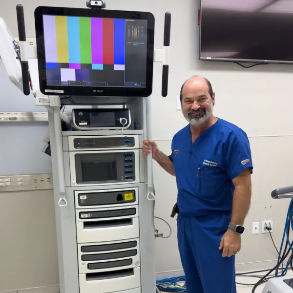
Dr. Brian Harkins is a renowned surgeon specializing in advanced, minimally invasive, and robotic surgical techniques. With a dedication to innovation and personalized patient care, he has transformed countless lives by delivering exceptional outcomes.
I want a website like this, where do i start?