Firefly Technology revolutionizes surgical precision through preoperative CT scan segmentation that generates detailed 3D spine renderings and personalized surgical plans. The system produces patient-specific autoclavable bone models and customized surgical guides via advanced 3D printing using medical-grade materials like PA12 nylon. These guides restrict drill paths to predetermined trajectories, achieving 99.7% accuracy in pedicle screw placement while reducing operative times and radiation exposure. The technology’s integration of CBCT imaging with specialized planning software guarantees optimal implant positioning and enhanced patient outcomes across complex spinal procedures.
While traditional spine surgery relies heavily on intraoperative imaging and surgeon experience, FIREFLY Guidance alters the surgical approach by shifting critical planning phases to the preoperative environment. This innovative workflow optimization begins with preoperative patient CT scans, which undergo segmentation to generate detailed 3D spine renderings. The system combines advanced imaging data with surgical planning software to develop personalized screw trajectories and implant plans through data driven surgical planning methodologies.
Digital plans are subsequently converted into physical models and guides via 3D printing technology, producing autoclavable, patient-specific bone models and surgical jigs. This predominantly preoperative workflow reduces intraoperative complexity and surgical time while supporting surgeon review and approval of surgical plans prior to surgery, ultimately enhancing precision and predictability. The system enables expert engineering support throughout the planning process, ensuring optimal outcomes for even the most complex spinal cases.
FIREFLY technology converts preoperative CT scan data into precise three-dimensional digital models that capture the patient’s unique anatomical structure. These models enable the creation of autoclavable bone replicas that surgeons can use for preoperative planning and procedural rehearsal. The system then fabricates customized surgical jigs with predetermined drilling angles and placement positions, ensuring optimal accuracy during the actual surgical procedure. The guides are manufactured using biocompatible materials that meet strict medical safety standards for surgical applications.
The integration of preoperative computed tomography data represents a cornerstone advancement in patient-specific 3D printed surgical guide technology. Cone Beam Computed Tomography (CBCT) serves as the primary imaging standard, capturing three-dimensional anatomical structures with voxel sizes ranging from 0.1 to 0.5 mm³. However, CBCT accuracy limitations of 200 to 1000 micrometers necessitate clinical safety margins of approximately 2 millimeters during guide design.
Enhanced precision emerges through multisensory data fusion, combining CBCT scans with intraoral scanner data that achieves superior accuracy of 20 to 100 micrometers for dental structures. This detailed 3D surgical planning approach compensates for CBCT’s lower dentition capture fidelity. Bilateral tooth-supported guides demonstrate superior accuracy compared to mucosa-supported alternatives, particularly in partially edentulous patients where enhanced stability contributes to optimal surgical outcomes. Radio-opaque markers facilitate precise correlation between imaging data and surgical guide positioning, ultimately enabling exact implant placement while preserving critical anatomical structures.
Beyond traditional imaging integration, patient-specific autoclavable bone models represent a pioneering advancement in surgical guide technology, enabling surgeons to practice complex procedures on exact anatomical replicas before entering the operating room. These models leverage medical-grade materials like PA12 nylon and specialized resin formulations that withstand standard autoclave cycles at 121°C or 134°C while maintaining dimensional accuracy. The manufacturing process requires careful material selection to meet regulatory compliance with biocompatibility standards. Studies demonstrate that proper material-printer combinations minimize post-sterilization deformation, preserving precise anatomical fit. Clinical outcomes show significantly improved pedicle screw placement accuracy and reduced operative times. Advanced SLS 3D printers like the EP-P3850 and EP-P420 provide the industrial-grade precision necessary for creating these critical medical components with consistent mechanical properties. This technology transforms surgical planning by providing tactile, sterilized models that enhance procedural precision and patient safety.
Advancing from anatomical modeling to functional surgical implementation, customized surgical jig fabrication converts patient-specific imaging data into precision-engineered guides that direct instrument placement during complex procedures. The digital workflow begins with CBCT and intraoral scanning to capture thorough anatomical details. Specialized planning software enables virtual implant placement while considering prosthetic requirements and anatomical constraints. The finalized design exports as an STL file for 3D printing using biocompatible resins through SLA or DLP technologies.
These patient-specific guides significantly enhance surgical precision by restricting drill paths to predetermined trajectories, minimizing deviation errors and anatomical damage risks. Clinical workflow optimization occurs through reduced surgical time and improved predictability. The technology enables prosthetic-driven planning that aligns implants with final restorations, ultimately providing increased patient satisfaction through enhanced outcomes, reduced invasiveness, and faster recovery times.
Precision in pedicle screw placement represents a critical determinant of surgical success in spinal procedures, where even millimeter deviations can result in catastrophic complications including spinal cord injury or vascular damage. Firefly Technology addresses this challenge by achieving 99.7% accuracy in pedicle screw placement through patient-specific 3D printed drill guides derived from preoperative CT scans. This exceptional precision reduces screw encroachment into vertebral canals while enabling optimal screw diameter and length selection before surgery. The system’s accuracy matches or surpasses existing piloted and robotic-assisted platforms while offering broader compatibility across screw systems. By minimizing placement errors, Firefly Technology delivers enhanced patient outcomes through reduced revision rates and improved construct stability, establishing a new standard for spinal surgical precision.
While surgical precision remains paramount, the radiation exposure associated with traditional intraoperative imaging presents significant health risks to both patients and surgical teams. Firefly technology addresses this concern through near-infrared fluorescence imaging, which reduces radiation exposure by up to 94% compared to conventional O-Arm systems. The technology employs indocyanine green dye to provide real-time visualization of vascular structures and tissue perfusion without ionizing radiation. This advancement enables effective radiation exposure mitigation while maintaining surgical accuracy. Integrated with robotic platforms, Firefly eliminates the need for frequent fluoroscopic verification during procedures. Intraoperative dose monitoring demonstrates substantially lower cumulative exposure for both patients and operating room staff, particularly benefiting vulnerable populations such as pediatric patients requiring multiple surgeries.
Fluorescence-guided surgery alters cancer detection by providing surgeons with real-time visualization of tumor tissues during operations. This technology enables precise identification of cancer margins and small tumor foci that remain invisible under conventional white light examination. The enhanced contrast between malignant and healthy tissues allows for more accurate tumor removal while preserving critical surrounding structures.
Cancer surgeons face a fundamental challenge in distinguishing malignant tissue from healthy structures during tumor removal procedures. Real-time fluorescence-guided surgery addresses this critical need by employing specialized contrast agents that selectively accumulate in cancerous tissues. When fluorescent dyes like indocyanine green or 5-aminolevulinic acid are introduced into the bloodstream, they concentrate within malignant cells, causing tumors to emit visible light under specialized cameras. Near-infrared imaging provides superior tissue penetration and spatial resolution for enhanced visualization. Multispectral imaging techniques further improve tumor detection by analyzing multiple wavelengths simultaneously. Ongoing research focuses on optimization of delivery routes to maximize dye uptake and minimize background fluorescence. This technology enables surgeons to achieve more complete tumor resection while preserving critical anatomical structures.
Achieving complete tumor removal requires surgeons to identify precisely where cancerous tissue ends and healthy tissue begins—a determination that can mean the difference between cure and recurrence. Fluorescence-guided imaging alters this critical assessment by providing real-time, high-resolution visualization of tumor boundaries during surgery. The technology distinguishes cancerous from normal tissue with unparalleled accuracy, detecting microscopic lesions that conventional methods often miss.
Clinical studies demonstrate sensitivity up to 100% and specificity up to 91% for detecting positive margins in head and neck cancers. This heightened precision enables flexible margin adjustment during intraoperative tumor staging, reducing reliance on subjective judgment. Back-table fluorescence imaging serves as a quality control measure, with results undergoing histopathological validation to certify accuracy and refine surgical techniques for optimal patient outcomes.
How can surgeons accurately determine tissue viability during complex procedures where visual assessment alone may prove insufficient? Firefly technology addresses this challenge through near-infrared fluorescence imaging that provides objective, real-time tissue viability assessment. When indocyanine green dye is administered, it fluoresces under NIRF illumination, creating quantifiable perfusion metrics that correlate directly with microvascular blood flow status.
This technology enables precise ischemic tissue detection by distinguishing between well-perfused and compromised tissues during reconstructive or resective surgeries. The fluorescence intensity and distribution patterns inform surgeons about tissue health with unparalleled accuracy. Profound tissue penetration capabilities allow assessment beyond superficial layers, supporting customized surgical approaches that preserve function. Clinical data demonstrate improved surgical safety and reduced complications through this objective viability monitoring, particularly in vulnerable structures where blood supply preservation remains paramount.
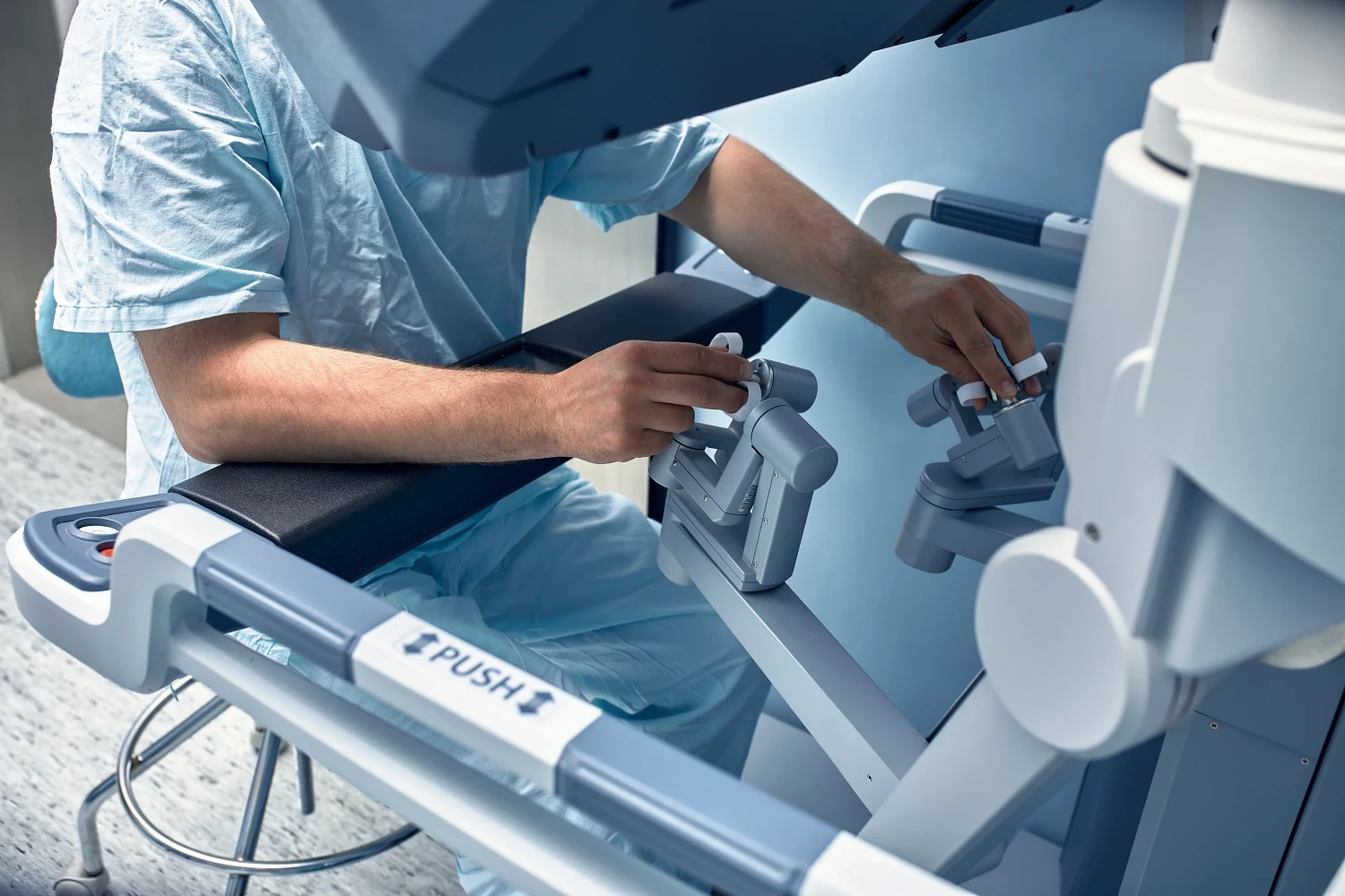
The seamless integration of Firefly technology within robotic surgical platforms represents a revolutionary shift in minimally invasive surgery, combining the precision of robotic manipulation with real-time fluorescence-guided visualization. This integration transforms surgical workflows by embedding near-infrared fluorescence imaging directly into robotic systems like the da Vinci XI, enabling surgeons to toggle between standard and fluorescence views without procedural disruption.
Enhanced surgical ergonomics emerge through synchronized controls that maintain natural workflow patterns while providing critical tissue perfusion data. The technology delivers improved visualization accuracy by offering magnified 3D fluorescence overlays that highlight vascular structures and tissue boundaries with unparalleled clarity. Surgeons can simultaneously view white light and fluorescence imaging, facilitating precise tumor resection while preserving healthy tissue, particularly in complex urologic and oncologic procedures requiring delicate anatomical guidance.
Multiple clinical studies demonstrate that Firefly technology delivers measurable improvements in surgical outcomes while streamlining operating room workflows. The enhanced visualization capabilities lead to improved patient outcomes through reduced anastomotic leakage rates and fewer postoperative complications. Studies report only minor grade 2 complications with no major adverse events documented in Firefly-assisted procedures.
Operationally, the technology integrates seamlessly with robotic systems, eliminating the need for separate ICG cameras and reducing setup complexity. Surgeons can switch between standard and fluorescence views without additional equipment, contributing to reduced operative time as proficiency develops. The streamlined workflow, combined with more precise tissue identification and vascular assessment, results in shorter hospital stays averaging 5.9 days and decreased reoperation rates.
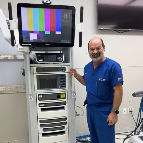
At Dr. Brian Harkins, we are proud to introduce FIREFLY technology — a significant advancement in surgical precision developed to support surgeons with enhanced visualization through fluorescence guidance and integrated navigation systems. FIREFLY delivers real-time tissue assessment, helps reduce radiation exposure, and improves screw placement accuracy, all of which contribute to measurable improvements in patient outcomes. As healthcare institutions increasingly adopt FIREFLY alongside robotic platforms, Dr. Brian Harkins positions this technology as an essential tool for modern surgical practice, delivering both clinical excellence and greater operational efficiency.
Robotic surgery offers enhanced dexterity, precision, and integration with firefly™ technology, unlike traditional laparoscopic surgery. While laparoscopy relies on rigid tools, the robotic surgical system provides a high-definition 3D view and smaller incisions, enabling minimally invasive procedures with reduced complications and faster recovery.
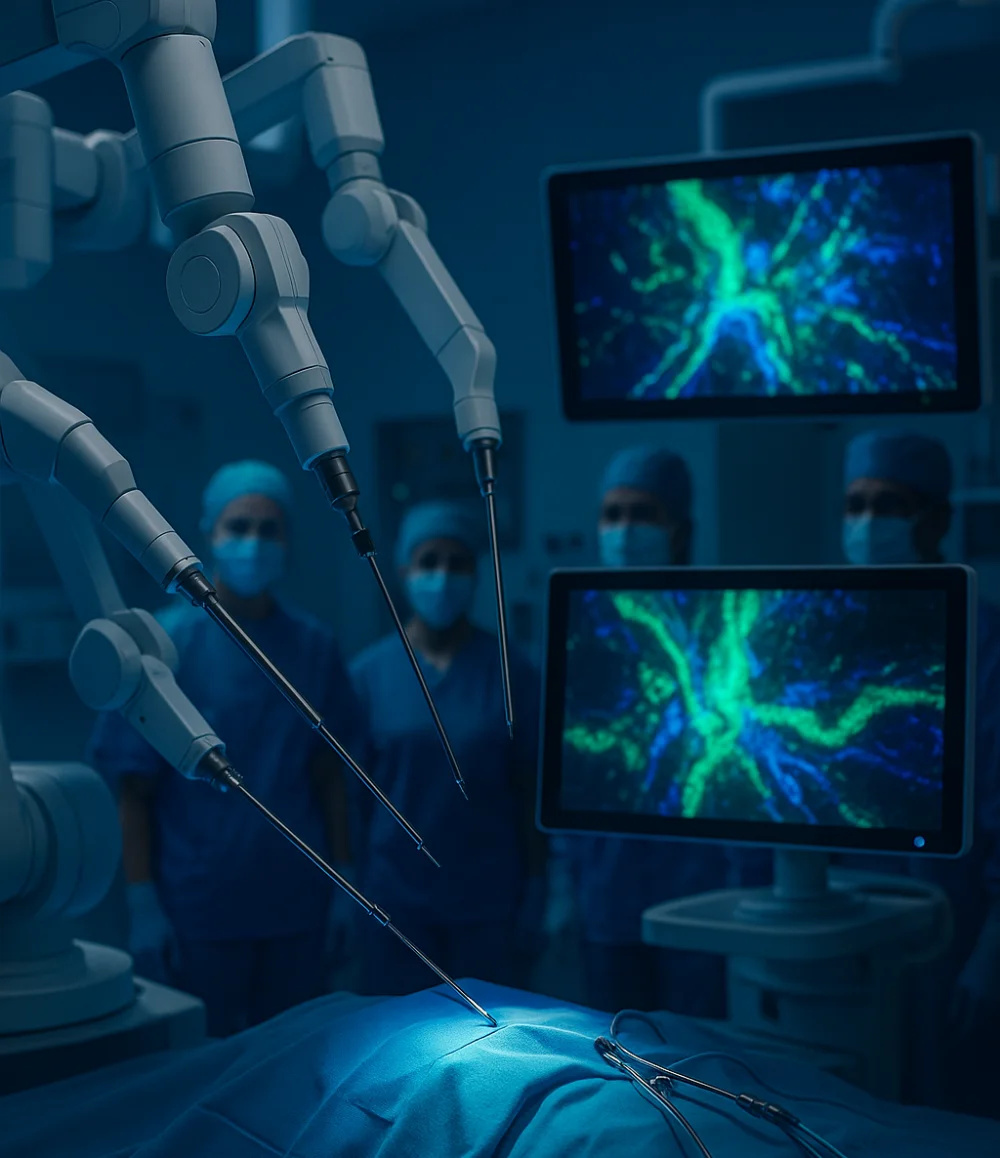
The firefly system integrated into the da Vinci surgical and da Vinci si surgical platforms enables fluorescence imaging capability that uses near-infrared light to highlight anatomy invisible to the naked eye. This technology makes it possible to differentiate blood vessels, ducts, and tissue structures with a distinct green glow, improving surgical safety.
In oncology and gynecologic surgery, firefly fluorescence imaging provides surgeons the ability to visualize tumor margins and distinguish the difference between cancerous and healthy tissue. This technology is particularly valuable in patients affected by endometriosis or those requiring ability to remove tumors without sacrificing the entire organ, ensuring more precise outcomes.
A robotic surgeon, operating the da Vinci surgical system, controls highly precise surgical instruments that work within the illuminated surgical field. The system enables surgeons to identify tissue planes and perform advanced surgical maneuvers with accuracy, reducing risks such as damage to the spinal cord during spinal surgery.
During a robot-assisted partial nephrectomy, firefly technology uses near-infrared imaging after dye is administered either intravenously to highlight the ureter and distinguish normal kidney tissue. This technique to identify perfusion patterns helps surgeons differentiate between cancerous areas and healthy structures, protecting ureteral anatomy and preserving function.
Firefly’s fluorescence imaging capability allows surgeons to make smaller incisions while still maintaining accurate anatomical orientation. This approach allows surgeons to spend less time in the operating room and reduces complications, showcasing how this new technique enhances recovery when used during robotic surgery.
In gynecologic procedures for patients affected by endometriosis, Firefly’s ability to visualize vascularized lesions within the surgical field makes resection more precise. The technology is particularly effective when combined with 3DHD optical imaging, which provides real-time identification of key anatomical landmarks that would otherwise be obscured.
In spinal surgery, Firefly’s optical integration provides a fluorescence imaging capability that aids in identifying vascular networks near the spine. This helps avoid damage to the spinal cord by improving the surgeon’s ability to visualize blood flow and anatomical planes, demonstrating how technology is a new safety standard in complex operations.
During hepatobiliary operations, Firefly fluorescence imaging capability is used during robotic surgery to visualize the common bile duct and common hepatic duct, reducing risks of accidental injury. The firefly technology uses near-infrared light to highlight these structures in a green glow, ensuring surgeons maintain accurate dissection planes.
Intuitive Surgical designs every medical device to integrate seamlessly into the da Vinci surgical system. Firefly allows the surgeon to combine fluorescence imaging in robotic-assisted surgery with laparoscopic efficiency, ensuring that surgery using this surgical system results in safer operations, reduced complications, and better recovery outcomes.
Dr. Brian Harkins is a renowned surgeon specializing in advanced, minimally invasive, and robotic surgical techniques. With a dedication to innovation and personalized patient care, he has transformed countless lives by delivering exceptional outcomes.
I want a website like this, where do i start?