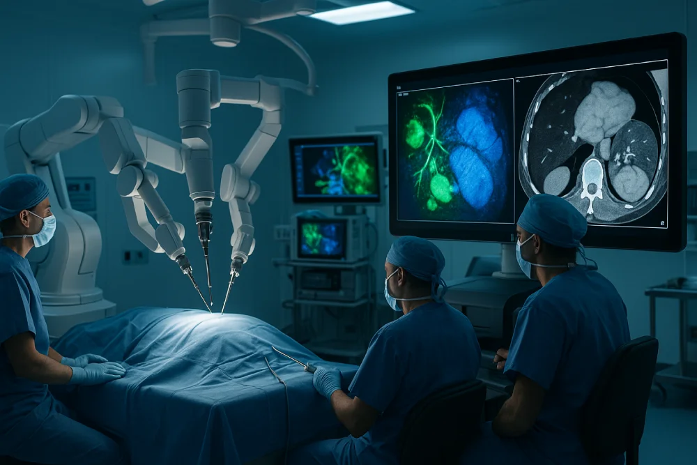
Advanced imaging systems have altered cancer surgery by providing real-time visualization of tumors and critical anatomical structures during operations. Near-infrared fluorescence imaging enables precise tumor margin identification using wavelengths between 700-900 nanometers for effective tissue penetration. Multimodal systems combine MRI, PET, CT, and optical imaging for extensive tumor assessment. C-arm CT integration supports minimally invasive interventions with 3D reconstruction capabilities. Intraoperative MRI compensates for brain shifts during procedures, maintaining surgical precision and reducing repeat surgeries. These technologies continue advancing surgical outcomes through enhanced precision.
Near-infrared fluorescence imaging has emerged as a revolutionary technology in cancer surgery, enabling real-time visualization of tumors and critical anatomical structures during operative procedures. This technique employs light wavelengths between 700-900 nanometers, which penetrate biological tissues effectively while providing excellent spatial resolution. Currently available imaging systems capture simultaneous visible and NIR fluorescence images, facilitating seamless surgical workflow integration without disrupting standard operative techniques.
The technology demonstrates particular strength in sentinel lymph node mapping, tumor margin identification, and assessment of tissue vascularization. ICG serves as the most widely-used fluorophore, though targeted contrast agents designed for enhanced tumor targeting specificity are under advanced clinical evaluation. These developments promise improved diagnostic accuracy and more precise tissue demarcation, ultimately supporting more conservative resections while reducing operative times and enhancing surgical outcomes.
Unlike traditional intraoperative imaging methods, near-infrared fluorescence imaging operates without ionizing radiation, making it an inherently safe option for repeated use during surgical procedures.
While near-infrared fluorescence imaging represents one powerful approach to intraoperative visualization, the broader panorama of molecular imaging encompasses a multifarious assortment of modalities that extend far beyond the surgical suite. Positron emission tomography remains the gold standard for whole-body cancer staging, utilizing radiolabeled tracers like ¹⁸F-FDG to visualize metabolic activity and enable quantitative tumor biology assessment. Emerging stimulated Raman scattering microscopy provides label-free, high-resolution imaging based on molecular vibrational signatures, with first-generation clinical systems now deployed for rapid intraoperative histology. Machine learning enabled imaging shows particular promise in automated tissue classification during surgery. Dynamic contrast-enhanced MRI offers perfusion data for tumor characterization, while optical molecular probes continue transforming toward enhanced selectivity and sensitivity for precise tumor margin delineation. Enzymatically activatable probes represent a particularly promising advancement, as these smart molecular agents can detect tumors both before and after surgical intervention by correlating contrast enhancement with actual tumor development.
As cancer surgery advances toward increasingly precise interventions, multimodal imaging systems have emerged as revolutionary platforms that synergistically combine multiple imaging modalities to enhance tumor detection, characterization, and real-time surgical guidance. The AMIGO suite exemplifies this integration, combining MRI, PET, CT, and optical imaging for thorough tumor visualization across brain, lung, prostate, and gynecologic cancers. These systems utilize complementary information—anatomical structure from MRI, molecular activity from PET, and metabolic changes from optical imaging—to improve surgical workflow optimization and precision margin definition.
Smart contrast agents, including superparamagnetic iron oxide nanoparticles, target specific molecular markers to enhance imaging specificity. Advanced tissue characterization methods, such as biomicroscopic systems combining ultrasound and multispectral imaging, enable rapid intraoperative assessment, ultimately reducing recurrence risk through complete tumor removal. The implementation of such systems requires a multidisciplinary team approach to effectively develop and integrate imaging, guidance, and therapy devices within the surgical environment.
Digital interventional imaging has altered cancer surgery by providing surgeons with enhanced visualization capabilities that enable precise tumor localization and real-time guidance during complex procedures. C-arm CT systems integrate fluoroscopy and computed tomography in mobile units, supplying high-resolution 3D imaging during operations. These systems support surgical workflow optimization by enabling continuous visualization of tumor boundaries, vascular structures, and surgical instruments throughout resection procedures.
The technology facilitates minimally invasive interventions while reducing blood loss and recovery times. Surgeons can make precise adjustments to their approach, minimizing healthy tissue damage and improving resection margins. However, perioperative radiation dose management remains critical to balance diagnostic benefits with patient safety. Advanced software integration enables 3D tumor reconstruction and augmented reality overlays, enhancing intraoperative steerage for personalized surgical strategies. These imaging systems address the need for dynamic real-time surgical guidance that traditional computer-assisted surgery technologies often lack.
How can surgeons achieve maximum tumor removal while preserving critical brain function during complex neurosurgical procedures? Intraoperative MRI (iMRI) provides real-time, high-resolution imaging that enables surgeons to visualize tumor margins fluidly during surgery. This technology compensates for brain shifts that occur during procedures, maintaining surgical precision throughout the operation.
| Capability | Clinical Impact | Outcome Benefit |
| Real-time imaging | Distinguishes tumor from healthy tissue | Improved extent of resection |
| Brain shift compensation | Maintains navigational accuracy | Reduced neurological deficits |
| Functional mapping integration | Preserves eloquent brain areas | Enhanced safety profile |
| Immediate feedback | Detects residual tumor tissue | Fewer repeat surgeries |
| High-field 3T systems | Superior image quality | Better surgical precision |
Studies demonstrate that iMRI leads to further tumor resection in 40% of cases, increasing gross total resection rates by over 30% while minimizing damage to critical brain structures.
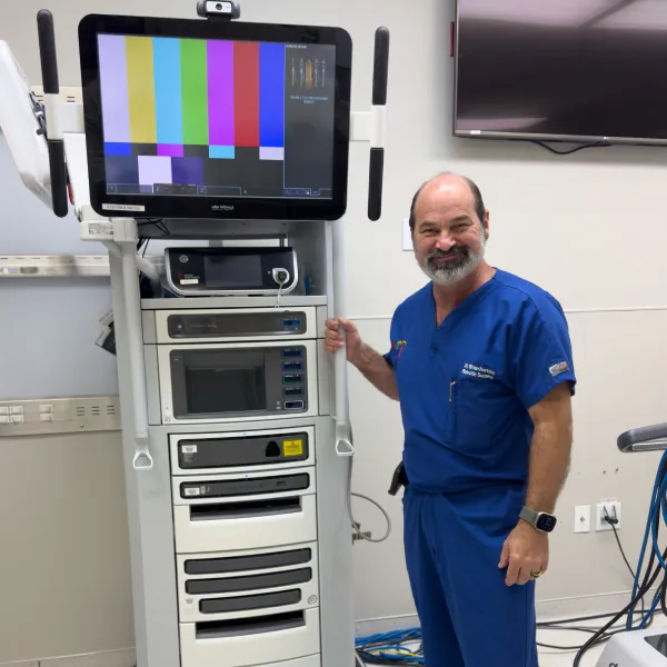
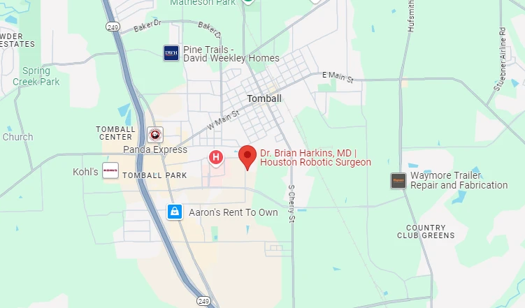
At Dr. Brian Harkins, we are advancing cancer surgery through state‑of‑the‑art imaging systems that enable real‑time tumor visualization and precise intraoperative guidance. Our use of near‑infrared fluorescence, targeted molecular imaging, and multimodal platforms provides surgeons with enhanced capabilities to distinguish malignant from healthy tissue. By integrating digital interventional imaging and intraoperative MRI into our surgical workflows, we improve accuracy in complex procedures such as brain tumor resection. These technological advances at Dr. Brian Harkins continue to optimize patient outcomes while minimizing complications in oncological surgery.
Modern imaging technologies provide clinicians with high-resolution medical images that improve the detection of cancer and support accurate cancer diagnosis. Techniques such as magnetic resonance imaging, ultrasound imaging, and photoacoustic imaging enable real-time visualization of cancer tissue and tumor cells, offering advantages over other imaging modalities for early cancer screening and effective diagnosis and treatment.
Image-guided surgery relies on different imaging modalities—including optical imaging systems, fluorescence imaging systems, and infrared fluorescence imaging—to direct surgeons during tumor resections. This approach ensures precise tumor removal while protecting healthy tissue, proving especially effective in breast cancer, prostate cancer, and colorectal cancer interventions, leading to better outcomes for cancer patients.
In breast-conserving surgery for breast cancer, surgeons use a fluorescence imaging system with indocyanine green fluorescence imaging for margin assessment in oral cancer and breast cancer sentinel lymph node biopsies. This image-guided cancer surgery enhances precision in breast-conserving surgery, reduces recurrence risks, and provides safer outcomes for patients with breast cancer.
Cancer research integrates biomedical imaging, optical imaging methods, and fluorescence molecular imaging to study tumor progression. Advanced imaging probes and imaging agents help visualize tumor cells at the molecular level, improving cancer diagnosis and treatment strategies. Publications like J Nucl Med Mol Imaging highlight how cancer using fluorescent imaging accelerates innovation in cancer imaging.
For head and neck cancer patients, advanced intraoperative near-infrared fluorescence imaging and fluorescence imaging and Raman spectroscopy allow precise image contrast and improved margin assessment in oral cancer. These imaging applications assist in surgery in head and neck, lowering recurrence rates and enhancing cancer treatment for oral cancer patients.
Intraoperative fluorescence imaging provides surgeons with real-time visualization of cancer tissue during minimally invasive surgery. Using dyes like indocyanine green fluorescence imaging, surgeons can perform precision surgery with clearer imaging of cancer boundaries in gastric cancer, pancreatic cancer, and lung cancer cases. This imaging approach reduces risks and improves recovery.
While standard imaging methods like ultrasound imaging and magnetic resonance imaging remain vital in cancer diagnosis, emerging tools such as optical imaging systems, infrared fluorescence imaging, and fluorescence molecular imaging offer advantages over other imaging modalities. These various imaging innovations provide improved imaging of glioblastoma, imaging of the gastrointestinal tract, and better oral cancer detection.
For biopsy in breast cancer and node biopsy in breast cancer, fluorescence-guided surgery with intraoperative near-infrared fluorescence imaging enhances accuracy. This technique improves image-guided identification of key lymphatic structures, ensuring surgeons achieve precise breast-conserving surgery while lowering recurrence rates for breast cancer patients.
In colorectal cancer and gastric cancer, surgeons rely on diagnostic imaging and image-guided surgery supported by fluorescence imaging systems to accurately identify tumor margins. These imaging techniques reduce complications, improve cancer treatment strategies, and enhance cancer diagnosis and treatment outcomes, particularly in precision surgery.
The combination of imaging and therapy creates a powerful framework for cancer diagnosis and treatment. Using imaging devices such as optical imaging systems, photoacoustic imaging, and intraoperative fluorescence imaging, surgeons tailor procedures to each patient. This integration ensures improved imaging of cancer, higher accuracy in cancer screening, and personalized treatment for conditions like oral cancer, ovarian cancer, and surgical hernia cases.
Dr. Brian Harkins is a renowned surgeon specializing in advanced, minimally invasive, and robotic surgical techniques. With a dedication to innovation and personalized patient care, he has transformed countless lives by delivering exceptional outcomes.
I want a website like this, where do i start?