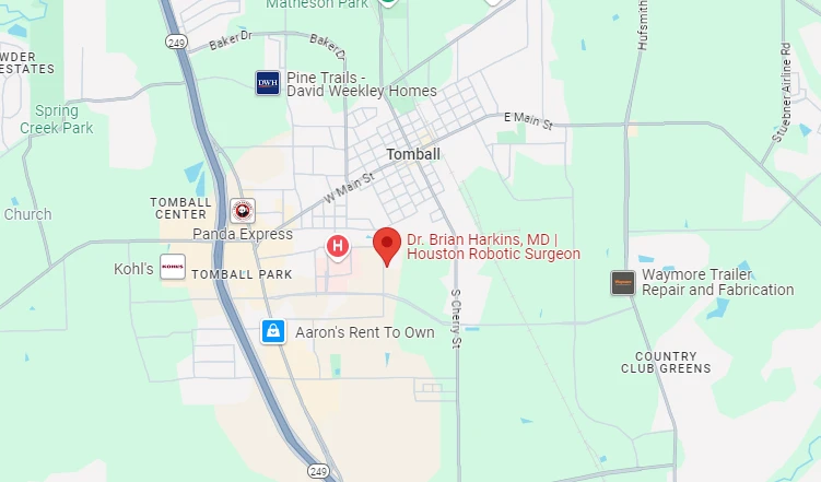
455 School St. Bldg. 1, Suite 10 Tomball, Texas 77375
Mon-Thu: 9:00 am – 5:00 pm | Fri: 9:00 am – 2:00 pm


Most hernias are confirmed by a physical exam alone, with about 95% of typical inguinal hernias in men diagnosed clinically without imaging. When the exam is inconclusive, ultrasound is usually ordered first, followed by CT for complex or post-surgical cases and MRI for occult hernias, where it offers 91% sensitivity and 92% specificity.
If you've noticed a bulge in your groin or abdomen that appears when you cough or lift something heavy, you might be wondering whether you have a hernia and what test can confirm it. Many people in Tomball, TX, are surprised to learn that most hernias are diagnosed through a simple physical exam by a surgeon, without needing imaging right away.
However, if your symptoms are unclear, you're overweight, or an ultrasound came back negative but you still feel something's wrong, additional tests like CT or MRI may be necessary. This article explains how doctors diagnose hernias, when imaging is actually needed, and which test is best for your situation.
A physical exam is the first step in confirming a hernia. During the exam, your doctor will inspect your groin or abdomen while you're at rest, then ask you to cough or bear down (called the Valsalva maneuver). This increases pressure in your abdomen and makes a hernia more visible or palpable.
For suspected inguinal hernias, the examiner may gently palpate the area with invaginated scrotal skin to feel for a bulge or impulse. They'll also check whether the hernia can be pushed back in (reducibility), which helps determine the type and severity.
Physical examination remains the preferred initial approach across clinical guidelines. It's quick, non-invasive, and highly accurate for typical cases, especially in men with straightforward inguinal hernias. National health resources such as MedlinePlus similarly describe hernias as commonly identified by the bulge they produce in the abdominal wall.
Physical exams aren't perfect in every situation. They can be less reliable in obese patients where subcutaneous fat obscures the exam, in people with surgical scarring, or when symptoms are vague without a visible bulge. In each of these cases the underlying hernia may be present but simply not palpable, which is why a normal exam alone does not always settle the question.
Hernia specialists and experienced surgeons tend to be more accurate diagnosticians than general practitioners, since they examine these presentations routinely and know which subtle findings matter. If your exam is inconclusive, your doctor will recommend imaging to confirm the diagnosis rather than leaving the question unresolved.
When the exam alone isn't enough, three imaging modalities are available, each with distinct strengths, timelines, and trade-offs.
| Test | Turnaround | Key Strength | Main Limitation |
| Ultrasound | About 30 minutes, often same or next day | Non-invasive, no radiation, real-time dynamic assessment | Highly operator-dependent; accuracy 33% to 86% for occult hernias |
| CT scan | Results in 24 to 48 hours | Comprehensive cross-sectional detail; less operator-dependent | Radiation exposure |
| MRI | Typically 1 to 2 weeks | Highest accuracy at 91% sensitivity and 92% specificity; no radiation | Longer wait times and higher cost |
Ultrasound is often the next step when a physical exam doesn't provide a clear answer. It's non-invasive, uses no radiation, and allows real-time, dynamic assessment as you move or strain. A standard hernia ultrasound takes about 30 minutes, and the technician can watch the area in motion, which helps identify hernias that only appear during activity or straining.
Ultrasound accuracy for occult hernias ranges from 33% to 86%, depending heavily on the operator's expertise. This high operator dependency means results can vary between facilities and technicians. A negative ultrasound does not rule out a hernia if clinical suspicion remains high, and false negatives are possible, especially for small or intermittent hernias. Ultrasound is recommended when the clinical exam is inconclusive and local ultrasound expertise is available.
CT scans provide comprehensive cross-sectional imaging that reveals complex anatomy and potential complications like bowel obstruction, strangulation, or mesh positioning from prior repairs. Results typically take 24 to 48 hours, and CT is less operator-dependent than ultrasound, making it more consistent across different facilities. However, it does expose patients to radiation.
CT scans are particularly valuable for post-surgical cases, recurrent hernias, and situations where detailed anatomical information is needed for surgical planning.
MRI offers the highest sensitivity (91%) and specificity (92%) for occult inguinal hernias. It can detect tears in abdominal muscles even when there's no visible bulge.
MRI is superior to ultrasound and CT for borderline cases where other tests have been inconclusive. The main drawbacks are longer wait times, typically 1 to 2 weeks depending on availability, and higher cost, but there's no radiation exposure. For patients with chronic groin pain and negative initial testing, MRI often provides the definitive answer needed to move forward with hernia treatment.
Not every hernia requires imaging beyond a physical exam. Your doctor will recommend imaging in specific situations:
These scenarios benefit from the additional detail that imaging provides, helping ensure accurate diagnosis and appropriate surgical planning. What they share is a gap between what the surgeon can feel and what is actually happening inside the abdominal wall, and imaging is the tool that closes that gap before any treatment decision is made.
The type of hernia and clinical situation determine which imaging test is most appropriate.
For an uncomplicated, symptomatic inguinal hernia in a man, the physical exam is often sufficient, and imaging can be skipped unless the presentation is atypical or the exam is uncertain.
When a hernia is suspected but not confirmed, ultrasound is typically ordered first due to lower cost and faster results. If the ultrasound is negative but clinical suspicion remains high, MRI is the next step.
Patients with prior hernia repair, mesh complications, or complex anatomy benefit from CT or MRI. These modalities provide the detailed anatomical information surgeons need to plan the repair safely.
Femoral hernias, more common in women, should be imaged due to higher strangulation risk. Ventral and incisional hernias also typically require ultrasound or CT to aid surgical planning.
The type of hernia influences which tests are needed. This overview of common hernia conditions helps explain why testing pathways differ.
Inguinal hernias in men are often diagnosable by physical exam alone, with about 95% of cases confirmed clinically without imaging. Femoral hernias, incisional hernias, and hiatal hernias are more likely to require imaging. Femoral hernias in women should always be imaged because they carry a higher risk of strangulation and require prompt surgical attention.
Different hernia types also have different appearances on imaging, which helps the surgeon understand exactly what needs to be repaired. That distinction matters because the imaging is not only confirming that a hernia exists, it is also defining its size, contents, and location so the repair can be planned around the actual anatomy rather than an assumption.
Understanding how long each test takes helps you plan your diagnosis and treatment. A physical exam takes just minutes during your office visit. Ultrasound takes approximately 30 minutes and is often available same-day or next-day. CT scan results come back in 24 to 48 hours, while MRI typically takes 1 to 2 weeks depending on facility availability.
Urgent hernias showing signs of strangulation, including severe pain, nausea, vomiting, or inability to reduce the bulge, bypass routine imaging and require emergency evaluation and possible immediate surgery. In those cases the priority shifts from confirming a diagnosis to protecting the trapped tissue, so waiting on a scheduled scan is not appropriate.
Three common presentations show how the testing pathway plays out in practice.
A 45-year-old man reports a groin bulge that appears with activity but causes no severe pain. During the physical exam, the doctor feels a palpable impulse in the groin when the patient coughs, and the hernia reduces easily when the patient lies down. In this case, the diagnosis is confirmed clinically without imaging, and surgery is scheduled electively, which aligns with how approximately 95% of male inguinal hernias are diagnosed.
A 50-year-old woman experiences chronic groin pain that worsens with exercise, but the physical exam reveals nothing. An ultrasound is ordered and shows posterior wall insufficiency but no discrete hernia. Because clinical suspicion remains high, an MRI is obtained, and it confirms a small direct inguinal hernia, allowing surgical planning to proceed. This demonstrates the step-wise imaging approach for occult cases.
A patient reports a bulge and heaviness two years after an open ventral hernia repair following bariatric surgery. A CT scan with contrast is ordered to assess hernia size, mesh position, loss of domain, and adhesions. The detailed findings guide the decision between laparoscopic repair and complex reconstruction, and this complex case justifies comprehensive imaging before any surgical intervention.
Hernia imaging costs vary by modality, and knowing the ranges helps you plan ahead.
| Imaging Test | Typical Cost Range |
| Ultrasound | $200 to $400 |
| CT scan | $800 to $1,500 |
| MRI | $1,000 to $3,000 |
Most insurance plans cover medically necessary imaging after a clinical exam, and Medicare generally covers hernia diagnostics when ordered appropriately.
Some insurance plans require imaging before approving a surgical consultation, while others cover only specific modalities. It's important to confirm coverage and pre-authorization requirements early to avoid delays in your diagnosis and treatment, since an unexpected denial can stall the process by weeks. If cost is a concern, discuss it with your doctor, who can help you understand which test provides the best value for your specific situation and work with your insurance to secure coverage.
Most hernias are confirmed by a physical exam, where your doctor inspects the area and asks you to cough or bear down. When the exam is inconclusive, imaging with ultrasound, CT, or MRI is used to confirm the diagnosis.
No. About 95% of typical inguinal hernias in men are confirmed clinically without imaging. Imaging is reserved for inconclusive exams, occult hernias, obesity, women with groin pain, recurrent hernias, and complex pre-operative planning.
MRI is the most accurate, offering 91% sensitivity and 92% specificity for occult inguinal hernias. It can detect tears in abdominal muscles even when no visible bulge is present, making it best for borderline cases.
Yes. Ultrasound accuracy for occult hernias ranges from 33% to 86% and depends heavily on operator expertise. A negative ultrasound does not rule out a hernia if clinical suspicion remains high, especially for small or intermittent hernias.
CT is used for complex anatomy, post-surgical cases, recurrent hernias, and suspected complications such as bowel obstruction, strangulation, or mesh positioning. It is less operator-dependent than ultrasound but does involve radiation exposure.
A physical exam takes minutes. Ultrasound takes about 30 minutes and is often available same-day or next-day. CT results come back in 24 to 48 hours, and MRI typically takes 1 to 2 weeks depending on facility availability.
Women face a higher risk of femoral hernias, which carry a greater strangulation risk and require prompt surgical attention. Femoral hernias in women should always be imaged rather than diagnosed on physical exam alone.
An occult hernia causes pain without a visible or palpable bulge. Because the physical exam may reveal nothing, these cases typically require imaging, starting with ultrasound and progressing to MRI if suspicion remains high.
Ultrasound typically costs $200 to $400, CT scans run $800 to $1,500, and MRI ranges from $1,000 to $3,000. Most insurance plans cover medically necessary imaging after a clinical exam, and Medicare generally covers appropriate diagnostics.
Seek emergency care immediately for severe pain, nausea, vomiting, or inability to push the bulge back in. These signs of strangulation bypass routine imaging and require urgent evaluation and possible immediate surgery.
For most people, the answer to what test confirms a hernia is simpler than expected: a careful physical exam by an experienced surgeon, which settles the great majority of straightforward cases without any imaging at all. Imaging earns its place when the picture is unclear, when body habitus or scarring limits the exam, or when the anatomy is complex enough that a surgeon needs to see it before planning a repair.
From there the path is stepwise. Ultrasound first for speed and cost, CT when detail and consistency matter, and MRI when suspicion persists despite negative results. Knowing which test fits your situation, and recognizing the red flags that call for emergency care instead, is what turns an uncertain symptom into a clear plan.
Get a clear answer about your symptoms.
Schedule an evaluation with a Tomball hernia specialist today.


Dr. Brian Harkins is a renowned surgeon specializing in advanced, minimally invasive, and robotic surgical techniques. With a dedication to innovation and personalized patient care, he has transformed countless lives by delivering exceptional outcomes.
I want a website like this, where do i start?