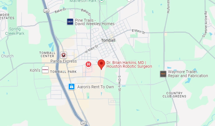
455 School St. Bldg. 1, Suite 10 Tomball, Texas 77375
Mon-Thu: 9:00 am – 5:00 pm | Fri: 9:00 am – 2:00 pm


Robotic umbilical hernia repair is a minimally invasive operation that closes the fascial defect at the navel and reinforces the repair with surgical mesh, performed through 3 to 4 small port incisions using the da Vinci Xi system. Most patients are discharged the same day or after one night, return to desk work in 1 to 2 weeks, and reach full activity at 4 to 6 weeks. Robotic repair shows particular advantage for adult umbilical hernias with diastasis recti, recurrent umbilical hernias, and hernias requiring mesh placement in the retromuscular plane.
The umbilical hernia is one of the most common types of ventral hernia in adults. It occurs at the navel, where a small natural opening in the linea alba (the central tendon line of the abdominal wall) can weaken over time and allow fat or intestinal contents to push through. By adulthood, the umbilical opening should have closed completely; when it has not, or when it reopens later in life, the result is an umbilical hernia.
Small asymptomatic umbilical hernias in adults can sometimes be observed safely. Symptomatic, enlarging, or complicated umbilical hernias usually need surgical repair, and modern repair is most often performed minimally invasively. Robotic assisted umbilical hernia repair, using the da Vinci Xi system, has expanded the range of cases that can be approached through small incisions, including hernias with coexisting diastasis recti, recurrent hernias, and hernias that benefit from retromuscular mesh placement. This guide walks through who needs surgery, what robotic umbilical hernia repair involves, what to expect during recovery, and how the diastasis recti question fits in. For patients in the Tomball area considering Dr. Brian Harkins, the procedure sits within his broader robotic surgery program at HCA Houston Healthcare Tomball.
An umbilical hernia is a defect in the abdominal wall fascia at or just below the navel. In infants, umbilical hernias are common and most close on their own by age 4 to 5. In adults, the umbilical opening should be fully closed; when it is not, or when an abdominal wall weakness develops at the navel later in life, the result is an adult umbilical hernia.
Risk factors for adult umbilical hernias generally fall into two categories: factors that increase intra-abdominal pressure, and factors that weaken the abdominal wall. Elevated BMI is among the most consistent risk factors because of the sustained increase in intra-abdominal pressure. Pregnancy, particularly multiple pregnancies, stretches the abdominal wall significantly and is a common contributor in women. Chronic heavy lifting (occupational or recreational), chronic cough, and chronic constipation all sustain elevated intra-abdominal pressure over time. Prior abdominal surgery near the navel can weaken the local fascia. Diastasis recti, the separation of the rectus muscles along the midline, often coexists with umbilical hernia and meaningfully influences repair planning. Rare connective tissue disorders also increase risk.
Umbilical hernias usually present as a soft bulge at or just below the navel, more prominent with standing, coughing, or straining. Symptoms range from cosmetic concern alone to mild dragging discomfort to significant pain with incarceration (contents stuck) or strangulation (blood supply cut off, a surgical emergency).
Surgical guidelines for adult umbilical hernia have shifted toward a more individualized approach. Small (under 1 to 2 cm), asymptomatic umbilical hernias in low risk patients can often be observed safely. Surgery is recommended when:
The decision is individualized. Hernias that are very small and asymptomatic may be safely watched. Hernias that are growing, symptomatic, or carry meaningful complication risk usually move to elective repair.
There are three modern approaches to umbilical hernia repair. All three are well established. The decision between them depends on hernia size, surgical history, anatomy, and surgeon experience:
For small primary umbilical hernias in healthy patients, all three approaches produce comparable outcomes. For mid sized hernias, hernias with coexisting diastasis recti, or hernias where retromuscular mesh placement is preferred, the robotic platform offers meaningful advantages.
Most adult patients with a symptomatic umbilical hernia are candidates for robotic repair. Specific factors that favor the robotic approach:
Patients who may not be candidates for minimally invasive repair include those with severe pulmonary or cardiac disease that contraindicates pneumoperitoneum, very large hernias requiring extensive component separation, severe coagulopathy, or specific anatomic limitations from extensive prior surgery.

Robotic umbilical hernia repair typically follows these steps:
Total operative time is typically 60 to 120 minutes for primary umbilical hernia repair, longer for combined repair with diastasis recti or for recurrent hernias.
Diastasis recti is the separation of the rectus abdominis muscles along the midline. It commonly coexists with umbilical hernia in adults, particularly in women after pregnancy and in patients with elevated BMI. The question of whether to repair the diastasis at the same time as the umbilical hernia depends on:
When diastasis is significant and symptomatic, robotic combined umbilical hernia and diastasis repair using techniques such as extended totally extraperitoneal (eTEP) repair allows both the hernia closure and the diastasis plication to be addressed through small port incisions, with mesh reinforcement in the retromuscular plane. This is an area where the robotic platform offers a meaningful advantage over open or laparoscopic repair.

Recovery from robotic umbilical hernia repair is faster than open repair and similar to laparoscopic. The general pattern:
Abdominal binder support for 2 to 4 weeks is commonly recommended after umbilical hernia repair to support healing and reduce post-operative seroma formation.
Published outcomes for robotic umbilical hernia repair in experienced hands are excellent:
Individual outcomes depend on hernia size, surgical technique, mesh selection, and patient factors.
Like any operation, robotic umbilical hernia repair carries risk, though most complications are minor and manageable. Bleeding or hematoma, surgical site infection, and seroma (a fluid collection at the surgical site that is usually self-limiting) are the most commonly discussed early complications. Mesh-related complications such as chronic pain, infection, or migration are rare but real, and the relevant risks vary by mesh product. Recurrence, bowel injury during dissection (very rare), the general risks of anesthesia, and the small possibility of conversion to open surgery should all be discussed during informed consent.
Most commercial insurance plans, Medicare, and Medicaid cover robotic umbilical hernia repair when medically indicated. Copays and deductibles are typically the same as for laparoscopic or open repair. Diastasis recti repair combined with hernia repair is usually covered when the hernia is the primary surgical indication. Standalone cosmetic diastasis repair is generally not covered. The billing team can verify your specific benefits before surgery is scheduled.
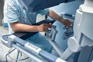
Robotic umbilical hernia repair is a minimally invasive operation that closes the fascial defect at the navel and reinforces the repair with surgical mesh, performed through 3 to 4 small port incisions using the da Vinci Xi platform. The surgeon sits at a console and controls every instrument movement; the robot translates those movements into precise actions inside the abdomen using magnified 3D vision and wristed instruments.
For small primary umbilical hernias in healthy patients, the three approaches produce comparable outcomes. For mid sized hernias, hernias with coexisting diastasis recti, or hernias where retromuscular mesh placement is preferred, the robotic platform offers meaningful advantages, including easier primary fascial closure and more precise mesh placement.
Surgery is recommended when the umbilical hernia causes pain or discomfort affecting daily activity, is enlarging over time, becomes incarcerated or strangulated, or carries meaningful complication risk. Small asymptomatic hernias in low risk patients can often be observed safely.
Yes. When diastasis recti is significant and symptomatic, robotic combined umbilical hernia and diastasis repair (often using extended totally extraperitoneal eTEP technique) allows both to be addressed through small port incisions, with mesh reinforcement in the retromuscular plane. This is an area where the robotic platform offers a meaningful advantage.
Most patients are discharged the same day or after one night, return to desk work in 1 to 2 weeks, drive within 3 to 5 days (off prescription opioids), and reach full activity at 4 to 6 weeks. Patients with combined diastasis recti repair or physically demanding jobs may need slightly longer.
For most adult umbilical hernias above approximately 2 cm in width, yes. Tissue only (no mesh) repair has historically had high recurrence rates. Mesh substantially reduces recurrence. Very small umbilical hernias (under 1 to 2 cm) may sometimes be repaired without mesh, but this decision is individualized.
Published 1 to 3 year recurrence rates are approximately 3 to 8 percent for primary umbilical hernia repair across robotic, laparoscopic, and open approaches in modern series. Lower for small primary hernias; higher for larger or recurrent hernias. Recurrence rates are also higher in patients with elevated BMI, diabetes, smoking, and chronic steroid use.
Mesh complications (chronic pain, infection, migration) are uncommon but real. The benefit of mesh in reducing recurrence substantially outweighs the small risk of mesh complications for most adult umbilical hernias. Mesh choice (synthetic, biologic, absorbable), weight, pore size, and placement strategy are individualized to the patient.
Yes, most primary robotic umbilical hernia repairs are performed as outpatient procedures, with same day discharge. Patients with larger hernias, combined diastasis repair, or significant comorbidities may stay one night for observation.
An umbilical hernia is a specific type of ventral hernia, located at the navel. The term "ventral hernia" is broader and includes umbilical, epigastric, spigelian, incisional, and parastomal hernias. Robotic umbilical hernia repair is essentially a focused subset of robotic ventral hernia repair, with the procedure adapted to the location and typical size of umbilical defects.
For adult umbilical hernia repair in 2026, patients have real options. Robotic umbilical hernia repair combines the precision of the da Vinci Xi platform with the recovery advantages of a minimally invasive approach, and is particularly well suited to mid sized hernias, hernias with coexisting diastasis recti, and cases where retromuscular mesh placement is preferred. For very small primary hernias, all three approaches (open, laparoscopic, robotic) produce comparable outcomes; surgeon experience and patient factors guide the decision.
If you would like to discuss robotic umbilical hernia repair for your specific case, schedule a consultation with Dr. Brian Harkins at our Tomball office by calling 281-351-5409 or request an appointment online. We see patients from across the greater Houston area, with surgeries performed at HCA Houston Healthcare Tomball's Center of Excellence in Robotic Surgery.
Sarah Chen is a healthcare communications specialist with 12+ years of experience in clinical environments. She writes about robotic and minimally invasive surgery for patients and families navigating surgical decisions. Her work is clinically reviewed by Dr. Brian Harkins, MD, FACS, ensuring medical accuracy and evidence-based information.
Based in: Tomball, TX | Serves: Greater Houston Area
This information is educational and is not a substitute for professional medical advice. Every patient is different. The decision to undergo robotic umbilical hernia repair is individualized based on hernia characteristics, anatomy, coexisting conditions (such as diastasis recti), comorbidities, prior surgical history, and patient preference. Dr. Brian Harkins will discuss whether robotic umbilical hernia repair is right for your specific situation in consultation. Statistics cited reflect published research on aggregate patient populations; individual outcomes vary. This content has been reviewed for medical accuracy by Dr. Brian Harkins, MD, FACS.

Dr. Brian Harkins is a renowned surgeon specializing in advanced, minimally invasive, and robotic surgical techniques. With a dedication to innovation and personalized patient care, he has transformed countless lives by delivering exceptional outcomes.
I want a website like this, where do i start?